
Arthrospira platensis vel Arthrospira maxima and Spirulina
Arthrospira platensis vel Arthrospira maxima is a gram negative, non-toxic species of cyanobacteria with a wide array of uses in the natural and commercial world. Cyanobacteria were earlier classified to group of so called blue-green microalgae. Arthrospira platensis has a characteristic blue-green colour (giving the prefix "cyano-") based on the wavelengths of light that it is able to absorb.
While many bacteria are known for their pathogenic effects, A. platensis is primarily known across the world for its potential nutritional value. It is one of the rare edible bacteria due to its low purine concentration, which allows it to pose very minimal risk of uric acid build up in the body. Historically, it is known to have been regularly consumed by the Aztecs and tropical climate populations where it was often dried into flat patties for easy consumption.
The consumption of this species has been shown to several beneficial health effects. The food industry classifies Arthrospira platensis as a single-celled protein, meaning that it is an edible microbe with a high food value. It is rich in vitamins, minerals, beta-carotene, essential fatty acids, and antioxidants, all of which have facilitated its commercial production as a human food supplement over the course of the past decade. It also has very high protein content with a well-balanced composition of all essential amino acids, making it even more desirable as a food supplement.
The commercial name of concentrated and lifilizated cyanobacteria Arthrospira platensis is SPIRULINA. This SPIRULINA is considered a "super food" by many scientists. According to a literature review researched by Cyanotech Corporation, Spirulina has "180% more calcium than whole milk, 670% more protein than tofu, 3100% more beta carotene than carrots, 5100% more iron than spinach, and more antioxidant and anti-inflammatory activity in 3 g of Spirulina than in five servings of fruits and vegetables".
Spirulina Extract
There are 3 types of spirulina extracts:
- Spirulina protein extract that shows excellent nutrional value and is considered as super food.
- Spirulina phycocyanin extract is classified as a colorant without an E number so that clear labeling is required.
- Spirulina lipoplysaccharide extract that shows immunomodulatory activity.
Phycocyanin belongs to group of anthocyanins – a group of naturally occurring pigments that are responsible for the red-blue color of many grains, fruits, and vegetables. Dietary consumption of anthocyanins is high compared with that of other flavonoids because of their wide distribution in plant foods. Numerous studies indicate the potential effect that this flavonoid family may have in reducing cardiovascular disease, cancer, hyperlipidemia, and other insulin resistance-related disease incidence through anthocyanin-rich food intake. The aim of the present chapter is to summarize known antidiabetic properties of plant-derived anthocyanins and anthocyanin-rich extracts to aid our understanding of their functional mechanisms. The chemical properties, dietary sources, metabolism, and bioavailability of anthocyanins will also be reviewed.
LCEPEEN Complex from Spirulina Extract
The Special Extract from Spirulina Platensis has been marked by the manufacturer as "LCEPEEN" registered code name. LCEPEEN is received in patented extraction process. LCEPEEN Extract is a novel immunomodulator derived from Spirulina platensis (Arthrospira), a member of the blue-green algae family (Cyanobacteria) . Preparations of Spirulina are popular dietary supplements that are consumed for their high nutritional value and their immunostimulatory properties. LCEPEEN Extract is a patented aqueous-alcohol extract of Spirulina containing high molecular weight polysaccharides. This preparation has been identified as a potent activator of cells of the immune system. In tissue culture, LCEPEEN Extract stimulates cells of the monocyte-macrophage system and dendritic cells to produce regulatory "pro-inflammatory" cytokines. In mice, LCEPEEN Extract is 10 times more efficacious than Spirulina in stimulating immune cells. In human monocytic THP-1 cells, this stimulatory effect of LCEPEEN Extract can be traced to the activation of NF-κB, a transcription factor that regulates the expression of genes involved in immune and inflammatory responses.
No toxic effects have been observed in cell culture or in laboratory animals. LCEPEEN Extract is safe in humans even when ingested in large amounts for extended periods of time. It has been used by many patients for nearly two years. It has efficacy at low doses after oral administration, and it is well tolerated. Clinical data collected so far show that LCEPEEN Extract provides an impressive range of health benefits in patients. It shortens the duration of infections, shows anti-viral activity in patients with herpes simplex-2 and HIV, reduces pain and strain, ameliorates swelling of inflamed joints and tissues, and accelerates wound healing. This broad range of health promoting effects distinguishes LCEPEEN Extract from other immunostimulants used clinically. The positive effect of LCEPEEN Extract on inflammatory pain and swelling of inflamed joints and tissues is a property not typically associated with immunostimulants. The biological and molecular mechanisms underlying these effects remain to be characterized. The clinical observations suggest that LCEPEEN Extract not only stimulates mechanisms involved in the initiation of inflammatory responses but can also modulate mechanisms involved in the resolution of inflammation. This unusual pharmacological profile suggests that LCEPEEN Extract has a positive effect on the overall response of the innate and adaptive immune system to foreign pathogens.
Uptake of High-molecular LCEPEEN Complex
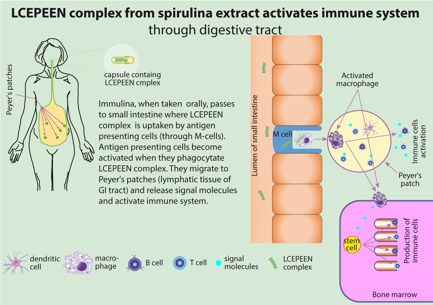
The molecule of LCEPEEN is too large to get absorbed from intestine. The LCEPEEN molecule is recognized by M-cells of lining mucosa of small intestine. M cells are structurally distinctive, uniquely permeable epithelial cells found only overlying the domes of mucosal lymphoid follicles. Antigenic macromolecules and some viruses, bacteria, and protozoa enter their apical surface by endocytosis or phagocytosis. These substances traverse the M-cell cytoplasm by transcytosis, breaching the epithelial barrier, and then interact with the subepithelial immunocompetent cells to initiate mucosal and systemic immune responses.
Through the M-cells antygens (like large molecules or patogens) can have further interactions with immune cells (antigen presenting cells: dendritic cells, macrophages, and B cells - so called professional Antigen Presenting Cells), which are found in the gut-associated lymphoid tissue (GALT) of the Peyer's patches in the small intestine, and in the mucosa-associated lymphoid tissue (MALT) of other parts of the gastrointestinal tract. Professional APCs specialize in presenting antigen to T cells. The main types of professional antigen-presenting cells are dendritic cells, macrophages and B cells. The professional APCs become activated when they phagocitate antigens. They start to release number of signal molecules (interleukines IL-1β, IL-6, IL-12) and migrate to other structures of the immune system like lymph nodes and bone marrow. Professional Antigen Presenting Cells are very efficient at internalizing antigens, either by phagocytosis (macrophages and dendritic cells) or by receptor-mediated endocytosis (B cells), processing the antigen into peptide fragments and then displaying those peptides, bound to a class II MHC molecule, on their membrane. The T cell recognizes and interacts with the antigen-class II MHC molecule complex on the membrane of the antigen-presenting cell. An additional co-stimulatory signal is then produced by the antigen-presenting cell, leading to activation of the T cell. The expression of co-stimulatory molecules and MHC class II are defining features of professional APCs. All professional APCs also express MHC class I molecules as well.
Biological Activity of Immulina in vitro
Immulina activates NF-κB in human monocytic THP-1 cells in vitro
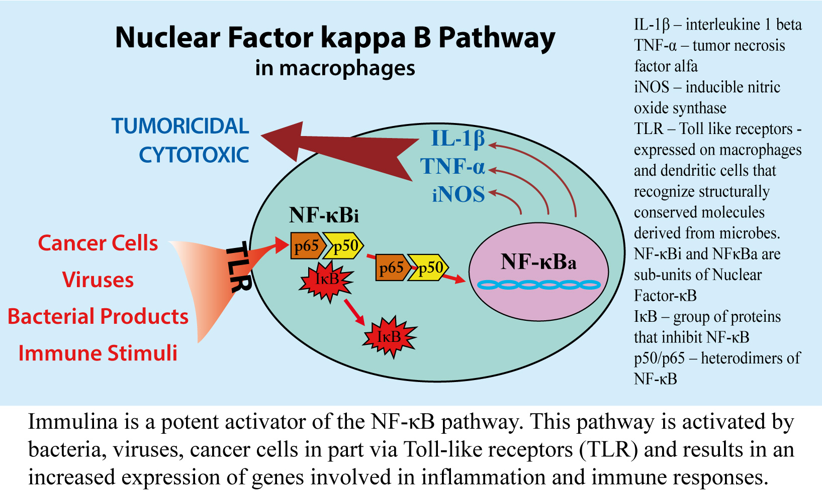
The innate immune system uses cytokines and chemokines in organizing and executing its response to foreign pathogens. The first step in this response is the activation of sentinel cells, which for the most part are macrophages. These cells are capable of recognizing many of the most common pathogens through specific receptors on their surface. A biochemical correlate of this activation is the rapid synthesis and release of cytokines and chemokines resulting in the recruitment of large numbers of monocytes and neutrophils from the circulation. Investigators at the University of Mississippi showed that this step is greatly facilitated by Immulina. They found that Immulina produces this effect by activating NF-κB (Figure 1). This transcription factor plays a pivitol role in mammalian innate immune responses since it can turn on genes that encode immune activators such as the cytokines tumor necrosis factor-alpha (TNF-α) and interleukin-1 beta (IL-1β), inducible nitric oxide synthase (iNOS), chemokines, adhesion molecules and cyclooxygenase-2 (COX-2). For their studies, these investigators used THP-1 cells, a human monocytic cell line that is commonly used as surrogate for macrophages and monocytes, the precursor cells of macrophages circulating in blood.

Investigators at the Johns Hopkins University described several genes encoding chemokines whose expression is enhanced in THP-1 cells after exposure to Immulina (Figure 3). These include the chemokines interleukin-8 (IL-8), mononuclear phagocyte chemoattractant protein-1 (MCP-1), macrophage inflammatory protein-1 (MIP-1) and interferon-gamma inducible protein-10 (IP-10).
Individual chemokines preferentially target different population of leukocytes. For example, IL-8 directs the recruitment of neutrophils from the circulation to sites of infections. Neutrophils make up 70% of the white blood cells in the circulation. Once they exit the vascular system, they become the principal killers of pathogens. MIP-1α and MIP-1β induce the influx of natural killer (NK) cells, macrophages and immature dendritic cells and IP-10 guides activated T cells into tissues.
Immulina activates dendritic cells in vitro
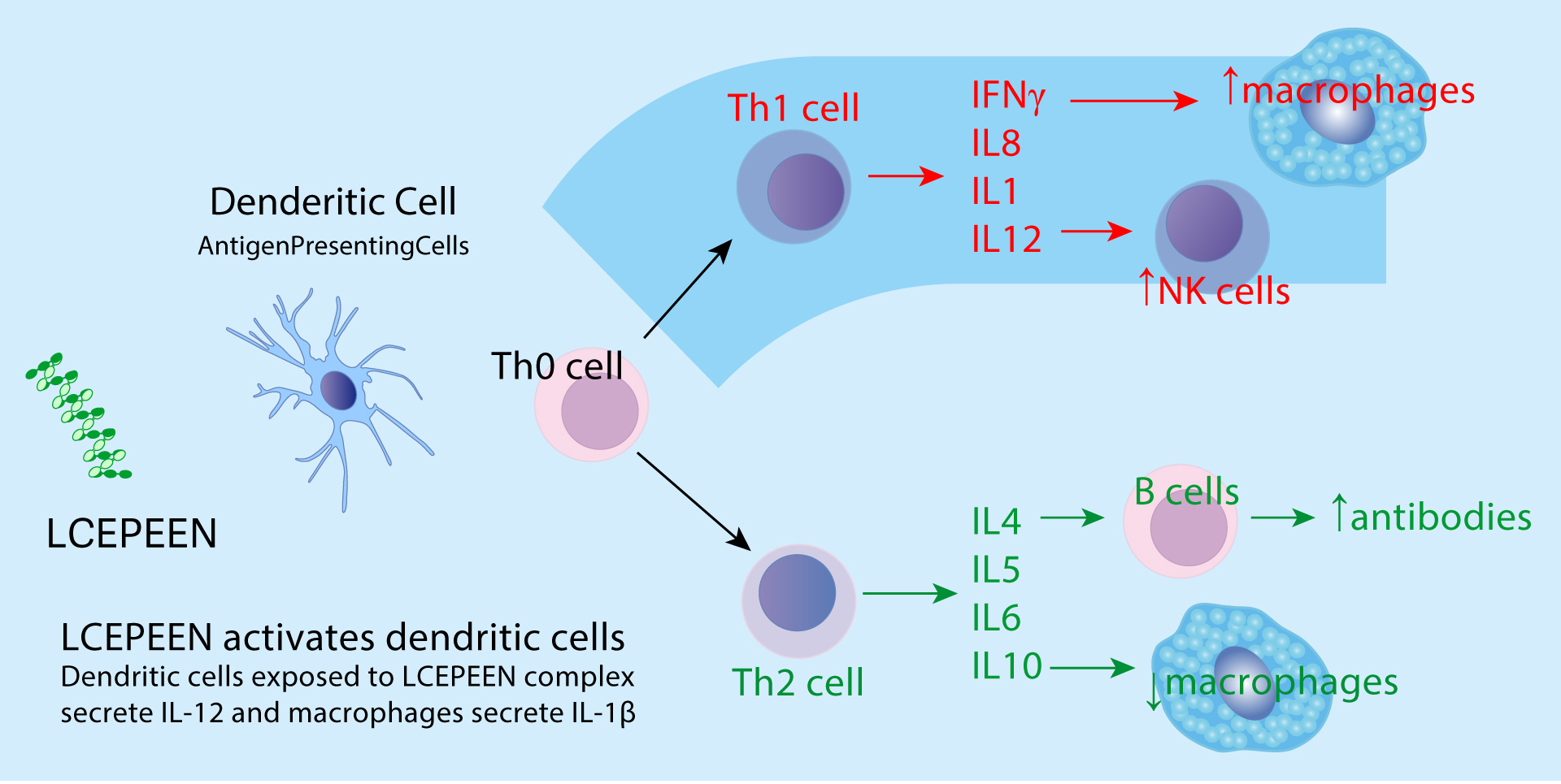
Investigators at the BioCentrum of the Technical University of Denmark found that Immulina can stimulate dendritic cells. These monocyte-derived cells are stationed beneath epithelial cell layers where they continuously sample the extracellular fluid for foreign antigens. When encountering bacterial products or increased cytokine levels, dendritic cells become activated and turn into antigen presenting cells. While macrophages remain stationary at sites of infection, dendritic cells migrate to lymph nodes. At these specialized lymphoid organs, dendritic cells present fragments of bacterial and viral proteins to naive T and B lymphocytes to initiate adaptive immune responses. The Danish investigators showed that murine dendritic cells when exposed to Immulina in culture for 20 hours, respond with an increased production of the cytokines IL-6, IL-10 and IL-12 (Figure 5). This observation indicates that Immulina can facilitate the transition of dendritic cells from antigen-capturing cells at sites of infection to antigen-presenting cells in lymph nodes. While still preliminary, this is an important finding since the types of cytokines secreted from dendritic cells determine the nature of the response of T cells in lymph nodes.
Immulina stimulates interleukin-12 (IL-12) production from peripheral blood mononuclear cells
An in vitro study at the Copenhagen University Hospital in Denmark found that Immulina at 2 μg/ml induces the production of IL-12 in human peripheral blood mononuclear cells. In contrast, IL-10 production was not affected. This finding indicates that similar to its action on monocytic cells, Immulina’s effect on peripheral blood mononuclear cells favors a Th1 immune response.